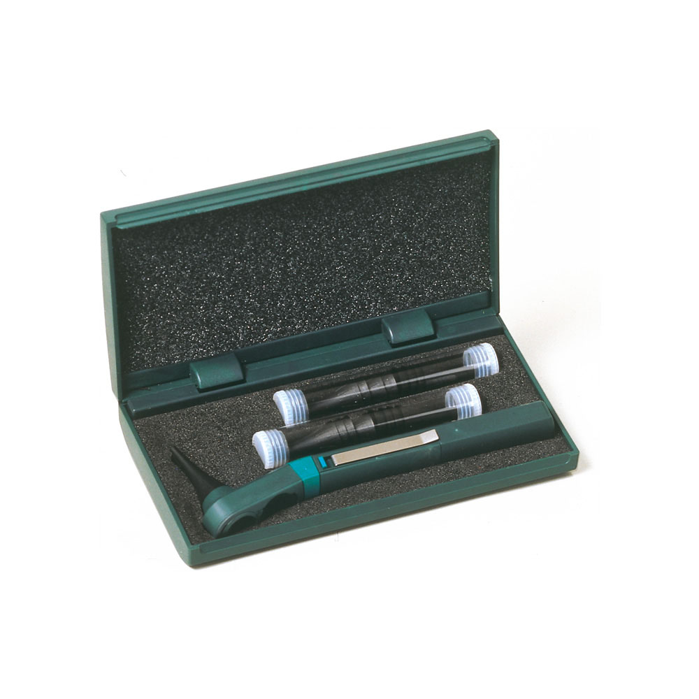
DISCONTINUED Digital Eye and Ear Examination Trainer Set, Dark
Adam Rouilly offers a wide selection of Clinical Skills Simulators - Ear/Eye Examination. Explore our diverse collection tailored for precise medical training needs. Our simulators provide effective tools for ear and eye examination techniques, meeting educational needs. Discover how our range enhances teaching and learning experiences.
See more detailsDiagnostic / Procedural Skills, Nursing, OSCEs, Paediatric Care, Physician Associates / PLAB / MLA, Postgraduate Clinical Skills, T-Levels, Trauma / Emergency / Acute Care, Undergraduate Clinical Skills.
Skills
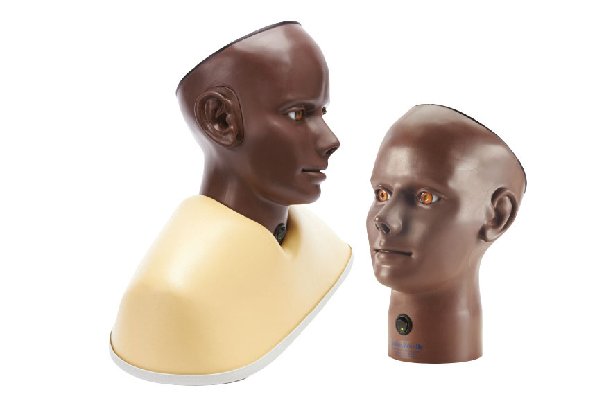
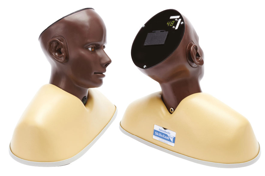
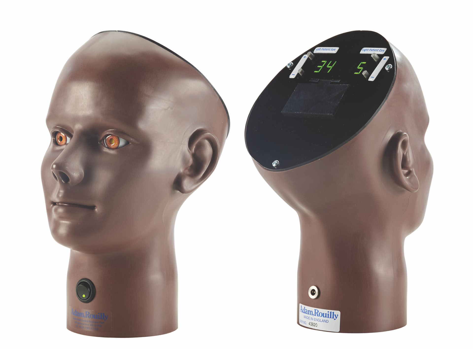
Adam,Rouilly’s new AR402-B DIGITAL EAR EXAMINATION TRAINER and popular AR403-B DIGITAL EYE RETINOPATHY TRAINER are now available together in an educational set ideal for any clinical skills setting.
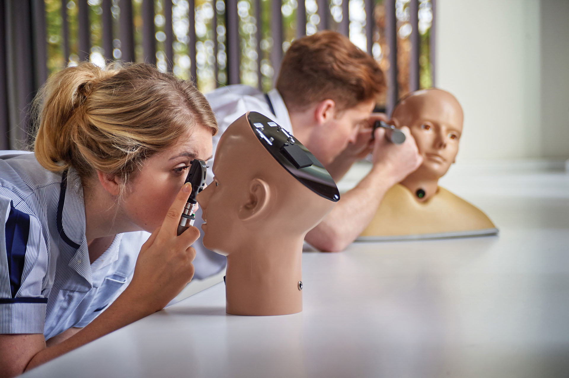
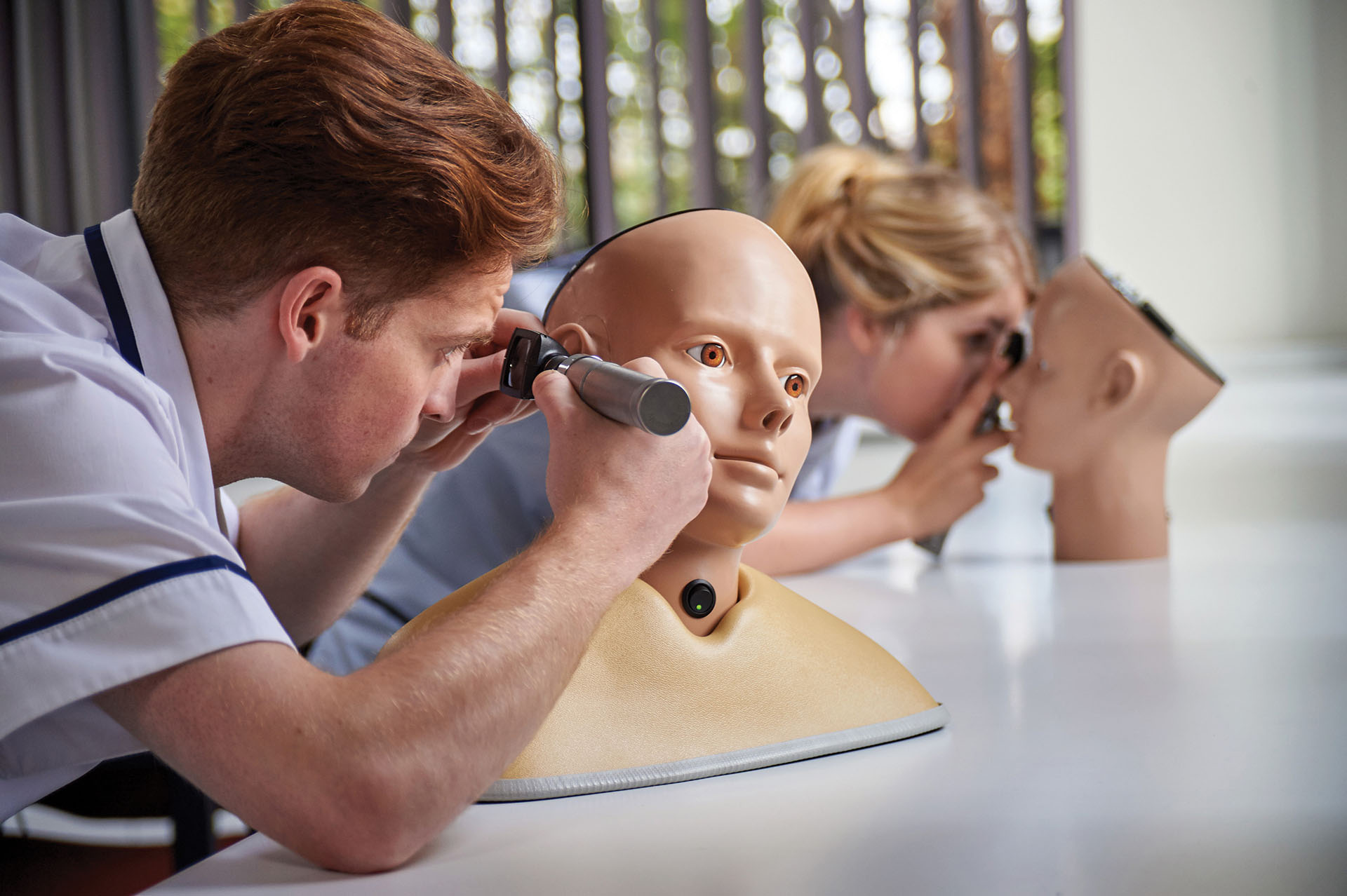
The AR402-B DIGITAL EAR EXAMINATION TRAINER features 48 ear conditions and the AR403-B DIGITAL EYE RETINOPATHY TRAINER features 36 eye conditions totalling 84 ear and eye conditions that form the ideal basis for both introductory and advanced ear and eye examination, as well as training in ophthalmoscope and otoscope use.
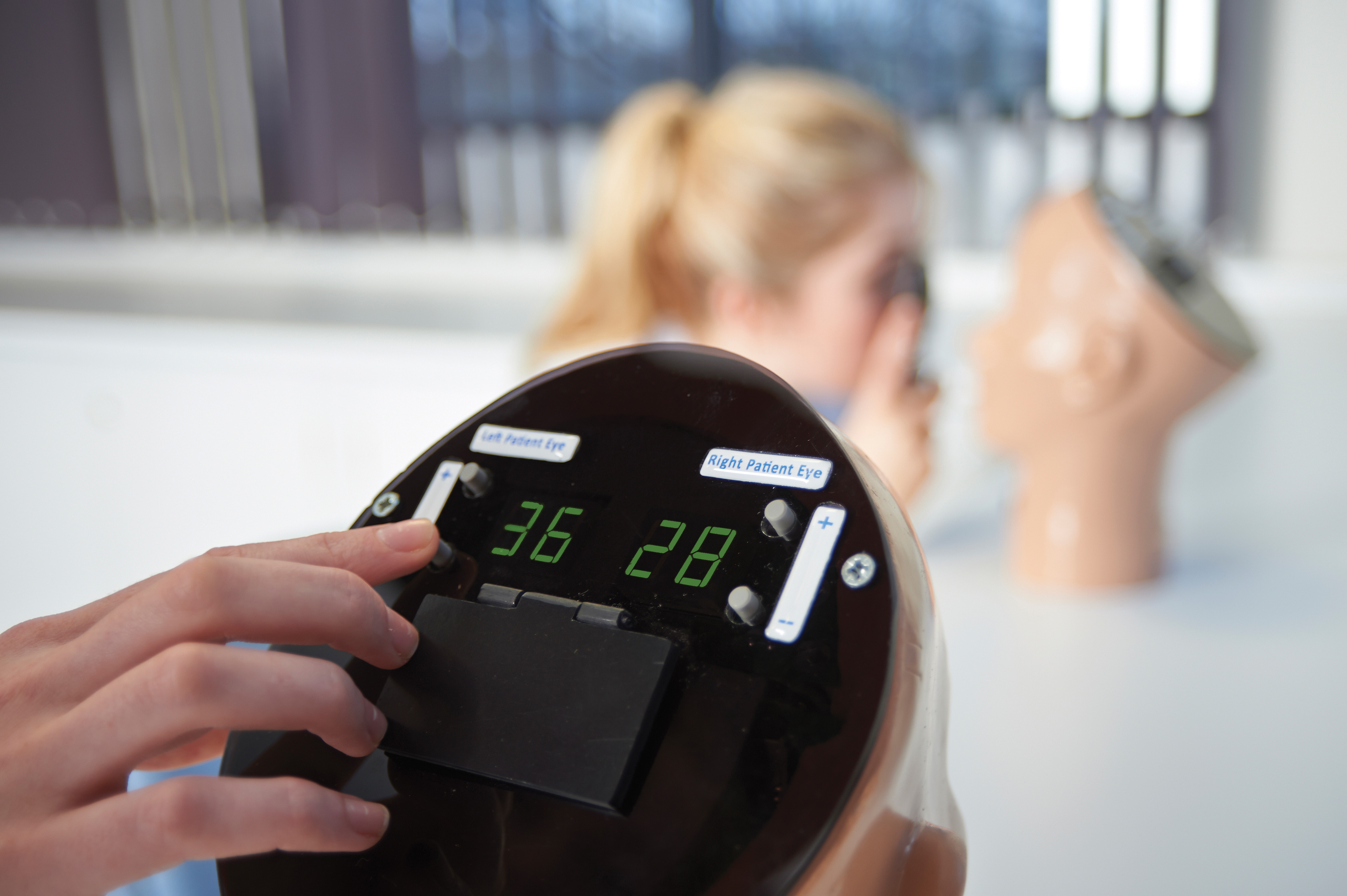
Both models feature high resolution displays on which each condition is represented, as well as life-size proportions and realistic features to greatly enhance any training scenario.
Features
- Cost saving advantage as a two model set
- Simple to set up and use
- 48 ear conditions
- 36 eye conditions
- High resolution digital displays
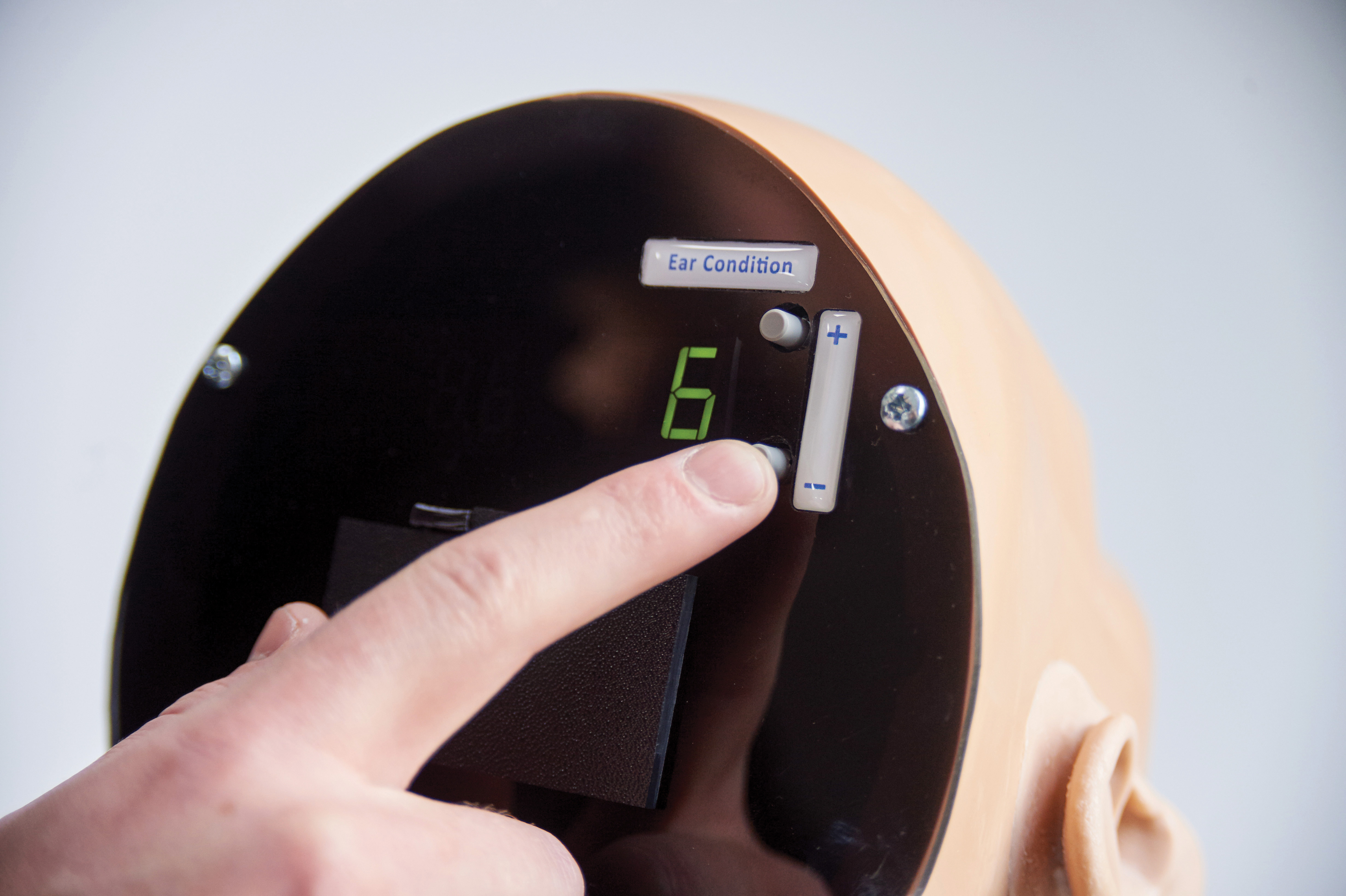
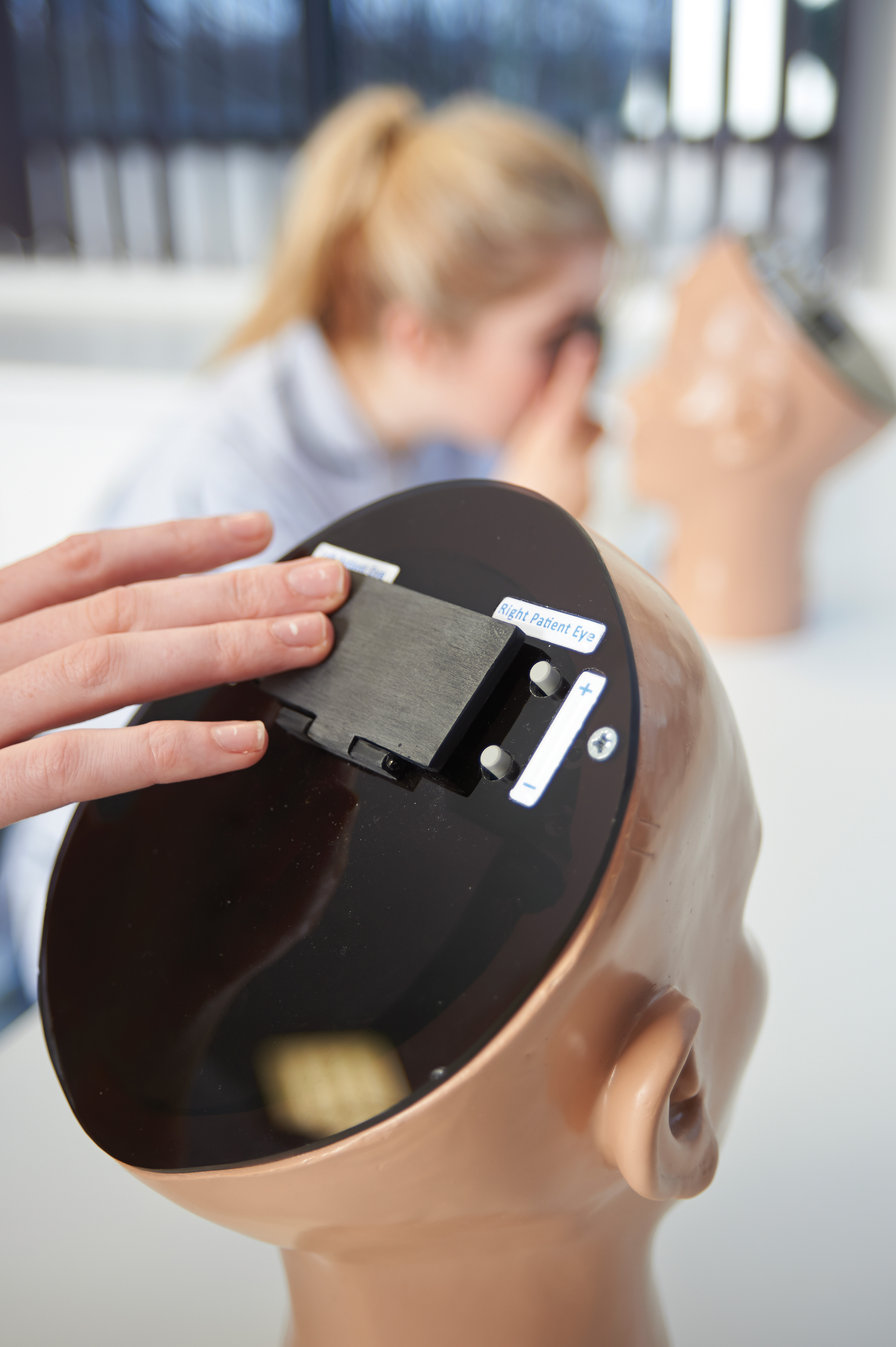
- Easy to use, digital control for eye and ear conditions
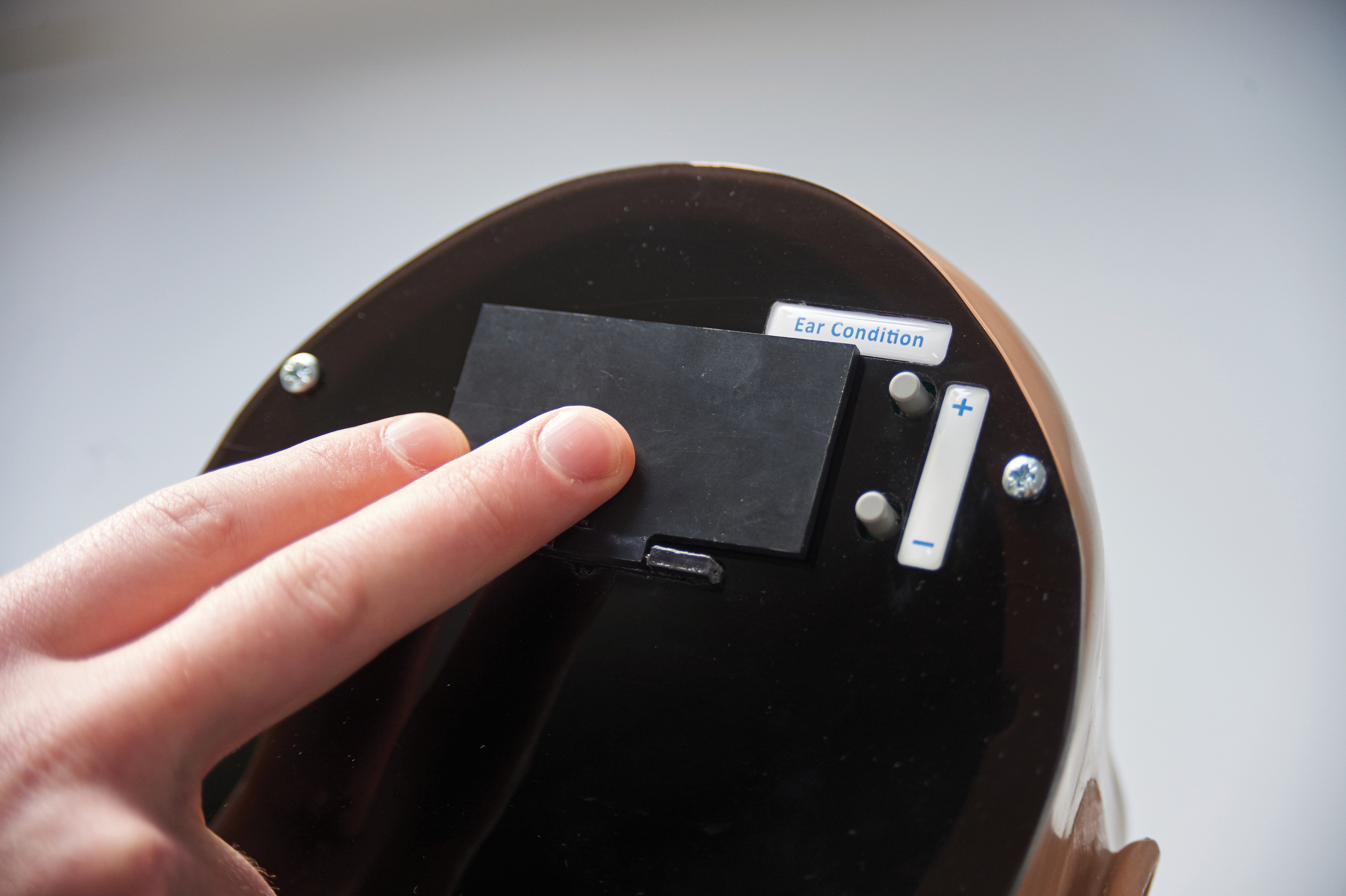
- Examination cover to hide displays of condition numbers
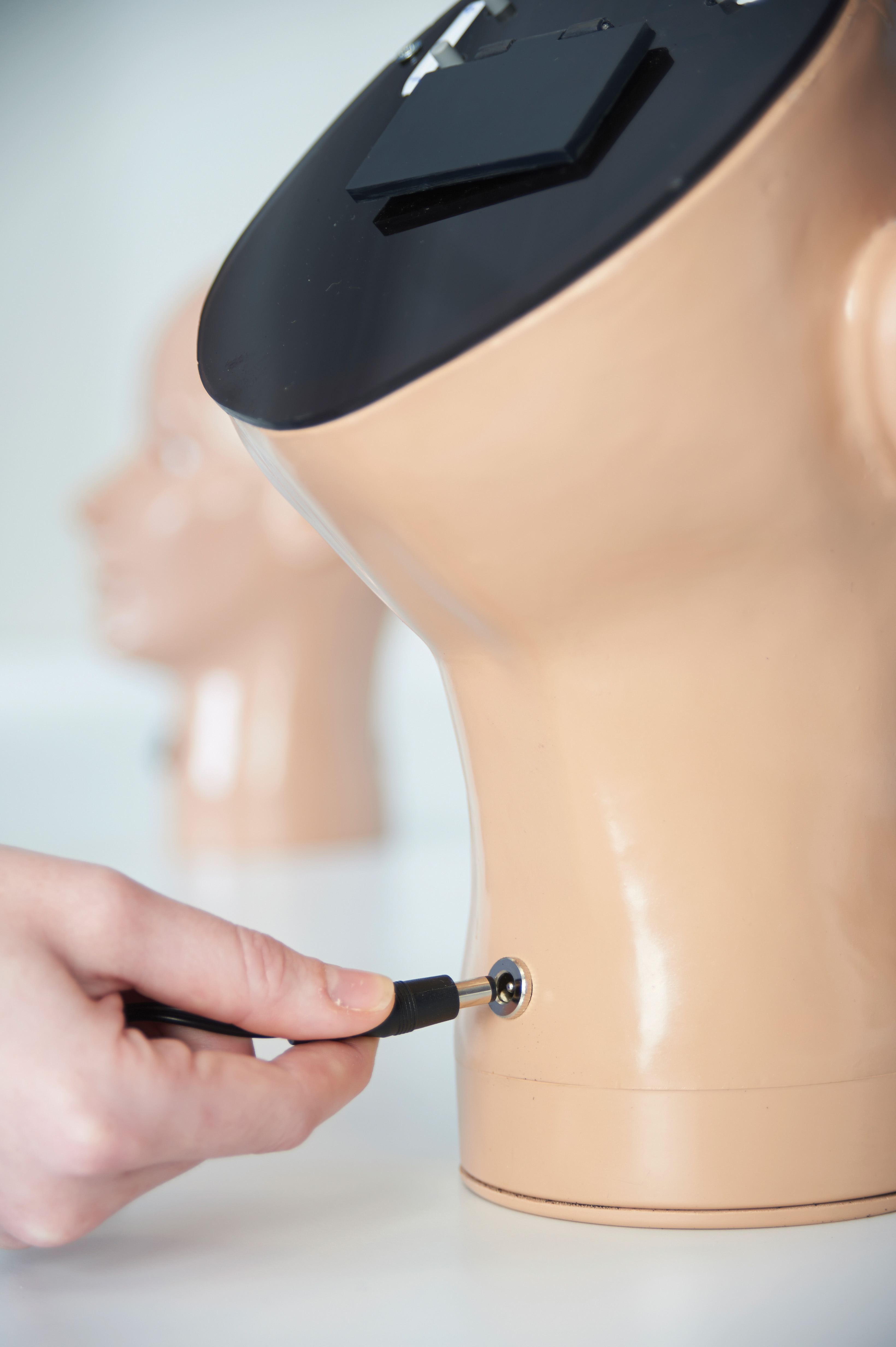
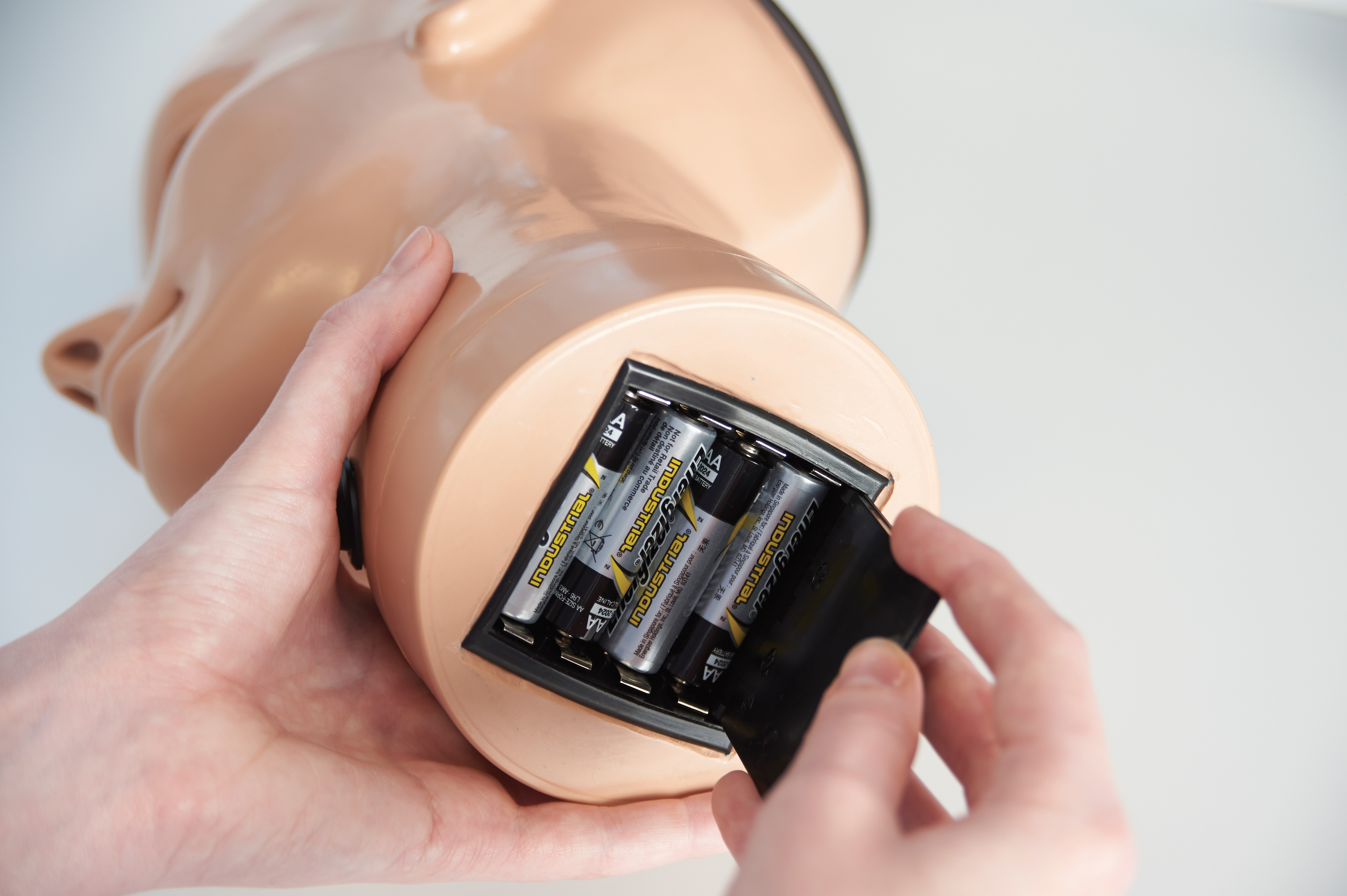
- Battery or worldwide mains power compatible
- Sleep mode to conserve power
2 Year Guarantee
- This model, manufactured by Adam,Rouilly, comes with a 2 Year Guarantee.
This guarantee applies to models which have been used correctly and covers durability and functionality.
Digital Ear Conditions
Ear conditions and diseases presented digitally for the right patient ear:
1. Normal I
2. Normal II
3. Ear Wax (Cerumen)
4. Swimmer’s Osteoma
5. Fungal Ear I
6. Fungal Ear II
7. Acute Viral Ear
8. Acute Secretory Otitis Media I
9. Resolving Secretory Otitis Media
10. Acute Secretory Otitis Media II
11. Acute Secretory Otitis Media III
12. Perforation following an Acute Suppurative Otitis Media (ASOM)
13. Childhood Glue Ear
14. Glue Ear in a Child with a Dermoid Cyst in the Eardrum
15. Adult Glue Ear
16. A Standard Ventilation Tube in the Membrane
17. Infected Mini Grommet with Otitis Externa Secondary to a Mucus Discharge
18. Permanent Ventilation Tube in Place
19. Large Perforation of the Tympanic Membrane
20. A Posterior Perforation of the Tympanic Membrane
21. Two Small Traumatic Perforations following a Blow to the Ear
22. Subtotal Perforation of the Tympanic Membrane
23. Perforation with Tympanosclerosis
24. Grommet Scar Healed
25. Tympanosclerosis of Tympanic Membrane
26. Posterior Retraction
27. Retraction onto Long Process of the Incus
28. Retraction with Loss of Long Process of the Incus and Keratin Trail
29. Retraction with loss of Long Process of the Incus
30. Posterior Retraction Pocket onto Jugular Bulb and with Middle Ear Fluid
31. Retraction with Early Keratin Build Up
32. Childhood Attic Retraction
33. Deep Attic Retraction
34. Attic retraction Accumulation Keratin – Underlying Cholesteatoma
35. Extensive Accumulation with Cholesteatoma in Middle Ear
36. Wet Cholesteatoma
37. Clean Dry Reconstructed Mastoid Cavity
38. Old Style Mastoid Cavity with Residual Cholesteatoma
39. Mastoid Cavity with Fistula into Lateral Semicircular Canal
40. Congenital Cholesteatoma
41. Large Congenital Cholesteatoma
42. Ear Canal Cholesteatoma I
43. Ear Canal Cholesteatoma II
44. Keratosis Obturans
45. Glomus Tympanicum Tumours
46. Glomus Jugulare Tumour
47. Foreign bodies
48. Aural Polyp
Digital Eye Conditions
Eye conditions and diseases presented digitally for left and right eye:
Diabetic Retinopathy
1. Background Diabetic Retinopathy
2. Maculopathy
3. Pre-Proliferative Diabetic Retinopathy
4. Proliferative Diabetic Retinopathy
5. New Vessels Disc
6. Laser
7. Photocoagulation
8. Ungradable
Important and Common Retinal Conditions
9. Normal
10. Glaucoma
11. Papilloedema
12. Optic Atrophy
13. Age-related Macular Degeneration (Dry)
14. Hypertensive Retinopahty
15. Central Retinal Vein Occlusion
16. Central Retinal Artery Occlusion
17. Drusen
18. Retinitis Pigmentosa
19. Medulated Nerve Fibres
20. High Myopia
21. Branch Retinal Vein Occlusion
Important and Less Common Retinal Conditions
22. Pre-Retinal Vein Occlusion
23. Multiple Retinal Haemorrhages
24. Retinal Detachment
25. Angioid Streaks
26. Benign Disc Neavus
27. Malignant Melanoma
28. Macular Haemorrhage
29. Choroidal Naevus
30. Macular Scar (Toxoplasma)
31. Cytomegalovirus Retinitis
32. Lipaemia Retinalis
33. Medusa Head
34. Myopic Crescent – Normal Choroidal Vessels
35. Sub Hyaloid Haemorrhage Resolving
36. Macular Burn
Includes
- Instruction manuals describing details of each condition
- Low voltage mains adaptors with worldwide plug fixings
- Removable Shoulder base for Ear Model
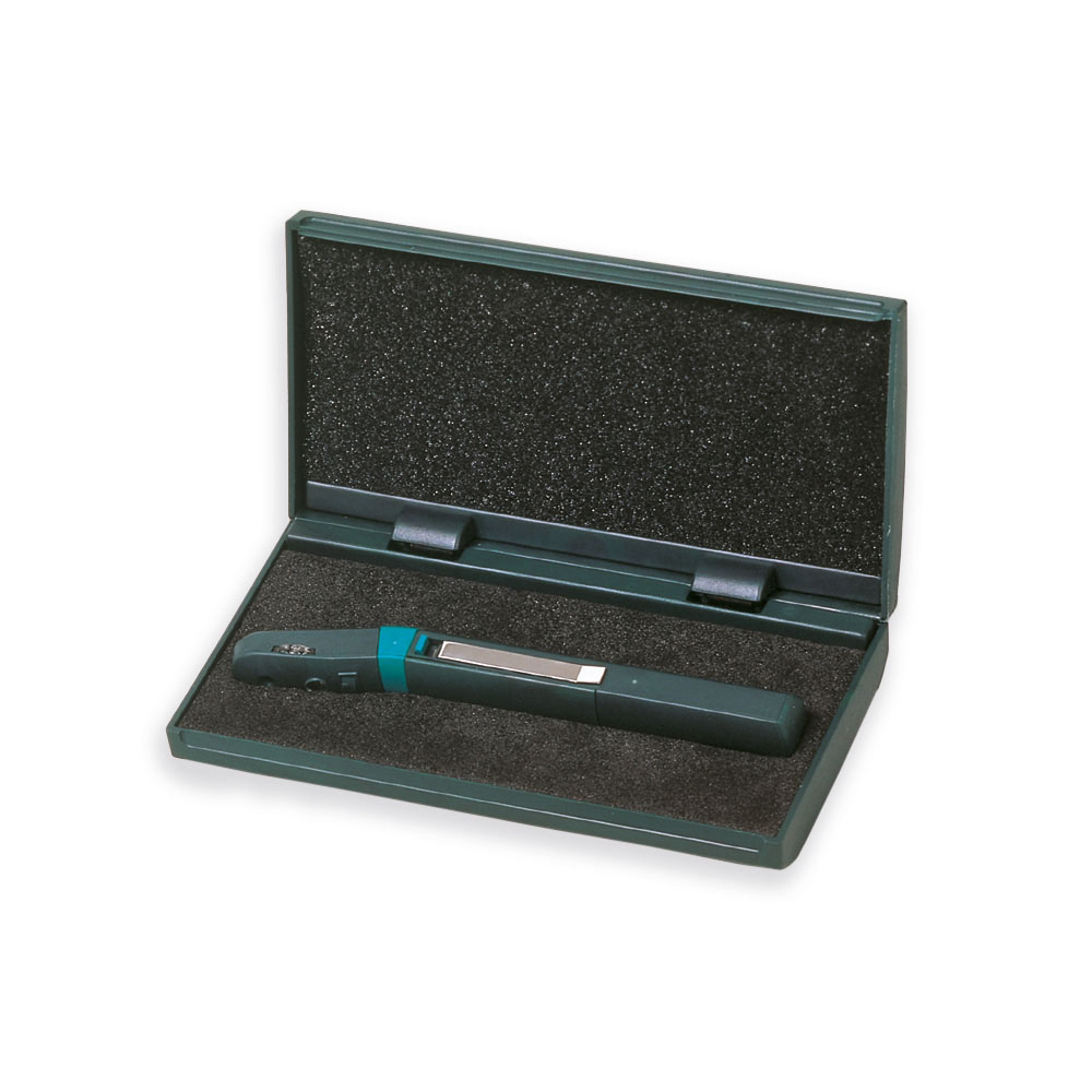
- Rigid carrying cases
Manufacturer
Adam,Rouilly
