
New
MO8/4
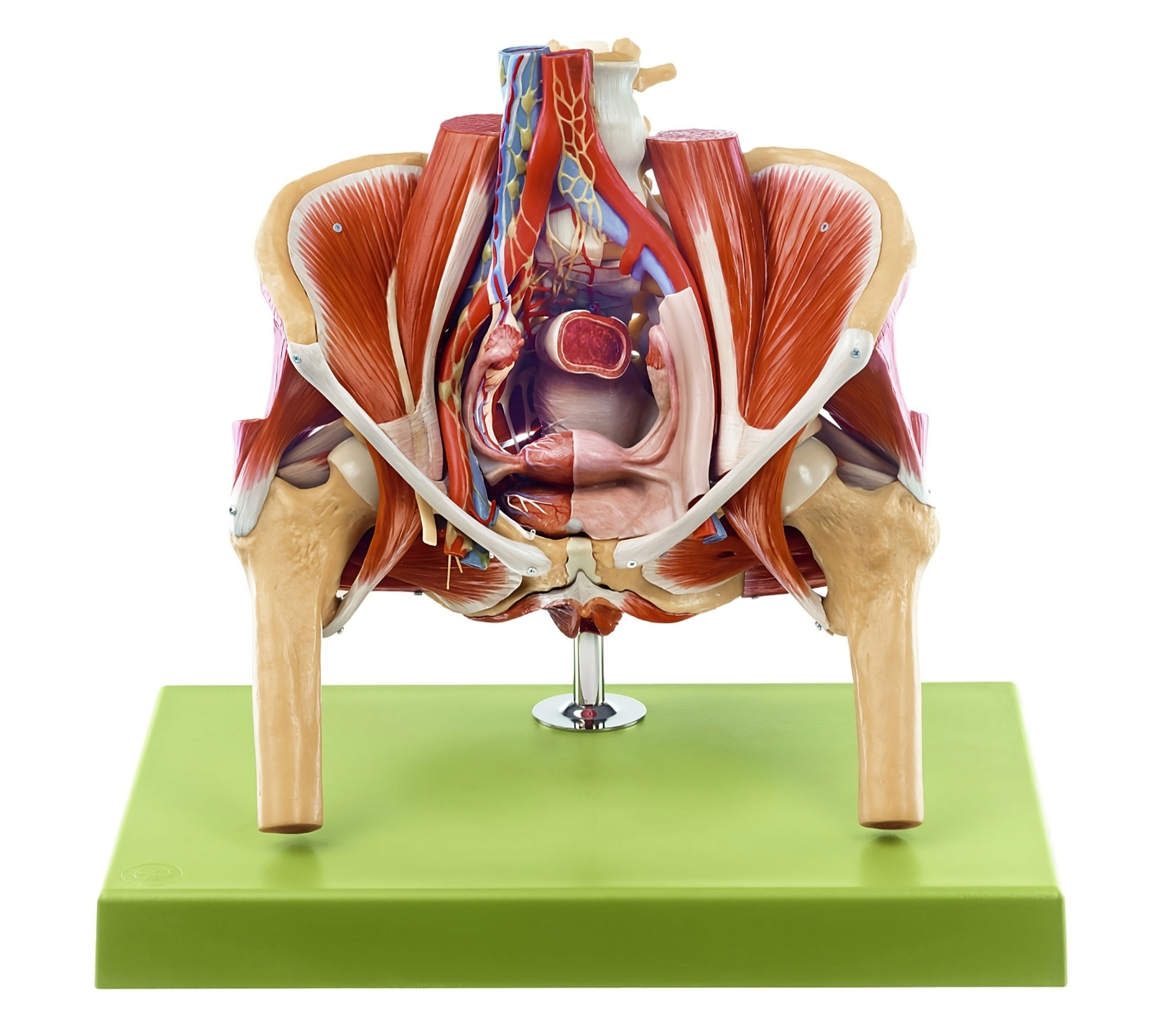
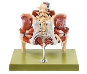
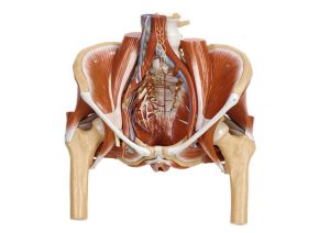
Model of the Female Pelvis
Male and female pelvis models showing pelvic bones, muscles, nerves, pelvic floor and reproductive organs for anatomy teaching. Ideal for anatomy teaching in medical schools, universities, colleges and secondary schools, from Adam,Rouilly, supporting education since 1918.
See more details
Price on request
Skills and Features
Features
- Natural size, in SOMSO-PLAST®
- After Prof. Dr. med.Elke Lütjen-Drecoll and Prof. Dr. med. Dr. med. hc. J.W. Rohen
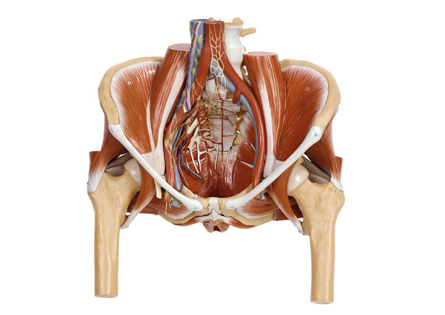
- Model of the female pelvis is a delicate model of the muscular floor of the pelvis which shows :
- Levator ani muscle with tendinous arch
- Urogenital hiatus (levator)
- Coccygeus muscle
- Internal obturator muscle
- Right side shows:
- The most important vascular, nerve, and lymphatic supply
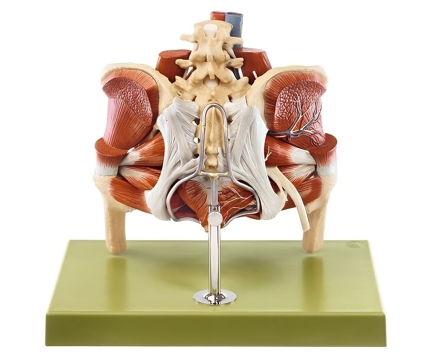
- Caudal view shows:
- Female pelvic floor with hip joints and symphysis
- Representation of the innervation of the external pelvic floor
- Cranial view shows:
- Female pelvis with tube and ovary
- Level of 4th lumbar vertebra
- Representation of the peritoneal relationships
- Separates into 4 parts including:
- Internal organs of the lesser pelvis with representation of the peritoneal relationships
- Lig. sacrotuberale
- External pelvic floor muscles
- On a stand with green base
- H 35 cm, W 39 cm, D26 cm
- Weight 4.2 kg
Additional Information
Manufacturer
SOMSO
