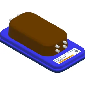
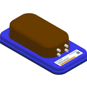
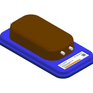
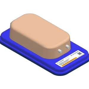
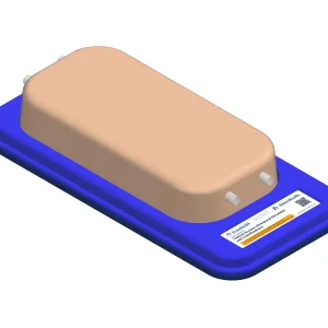
| Description | Designed for realistic, repeatable ultrasound-guided vascular access training, this simulator supports probe control, anatomy recognition and invasive technique practice with minimal setup. Lifelike echogenic material mimics human tissue, while two bifurcated femoral veins (approximately 8 mm) positioned at around 20 mm depth allow accurate needle insertion and ultrasound confirmation. | Designed for realistic, repeatable ultrasound-guided vascular access training, this simulator supports probe control, anatomy recognition and invasive technique practice with minimal setup. Lifelike echogenic material closely mimics human tissue, while four central veins (8–10 mm) positioned at approximately 15 mm and 35 mm depth allow accurate needle insertion and confirmation. | Designed for realistic, repeatable ultrasound-guided vascular access training, this simulator supports probe control, anatomy recognition and invasive technique practice with minimal setup. Lifelike echogenic material mimics human tissue, while two branched veins (approximately 8 mm, 10 mm and 20 mm) positioned at around 20 mm depth enable accurate needle placement and ultrasound confirmation. | Designed for realistic, repeatable ultrasound-guided vascular access training, this simulator supports probe control, anatomy recognition and invasive technique practice with minimal setup. Lifelike echogenic material mimics human tissue, while two bifurcated femoral veins (approximately 8 mm) positioned at around 20 mm depth allow accurate needle insertion and ultrasound confirmation. | Designed for realistic, repeatable central venous access training, this simulator supports probe control, anatomy recognition, and invasive technique practice with minimal setup. Lifelike echogenic material mimics human tissue, while two central veins (8 mm and 10 mm) positioned approximately 20 mm deep allow accurate needle insertion and ultrasound confirmation. | Designed for realistic, repeatable ultrasound-guided vascular access training, this simulator supports probe control, anatomy recognition and invasive technique practice with minimal setup. Lifelike echogenic material mimics human tissue, while two branched veins (approximately 8 mm, 10 mm and 20 mm) positioned at around 20 mm depth enable accurate needle placement and ultrasound confirmation. | Designed for realistic, repeatable Seldinger technique training, this simulator supports probe control, anatomy recognition, and vascular access practice with minimal setup. Lifelike echogenic material mimics human tissue, while two veins (8 mm and 10 mm) positioned approximately 25 mm deep allow accurate needle insertion and ultrasound confirmation. | Designed for realistic central venous access training, this simulator features two veins with simulated calcifications (approx. 10 mm diameter) for accurate needle insertion and assessment. Lifelike echogenic material mimics human tissue, supporting probe control, anatomy recognition, and safe practice of ultrasound-guided invasive techniques in a controlled, repeatable learning environment. | |